A 2-year-old boy presents with asymptomatic keratotic papules on his hands and feet. A biopsy is performed and he is confirmed to have punctate porokeratotic keratoderma. A discussion of punctate porokeratotic keratoderma is provided along with treatment options.
|
|
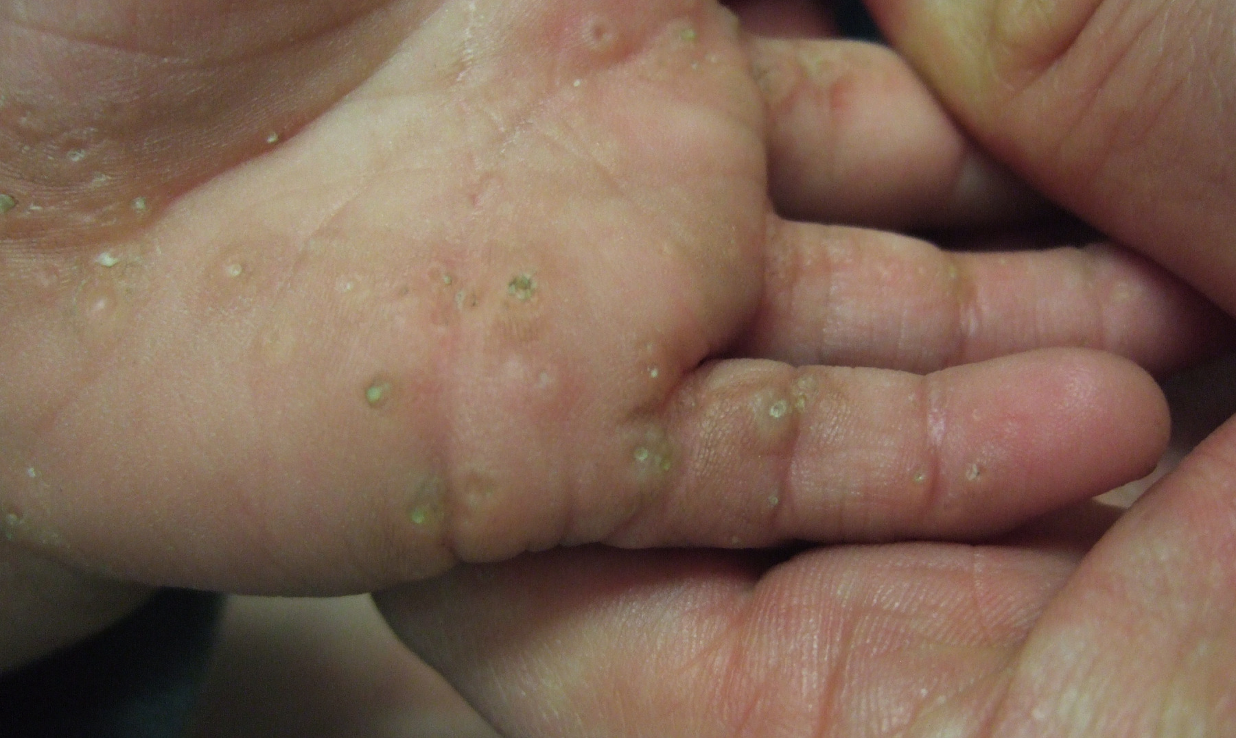
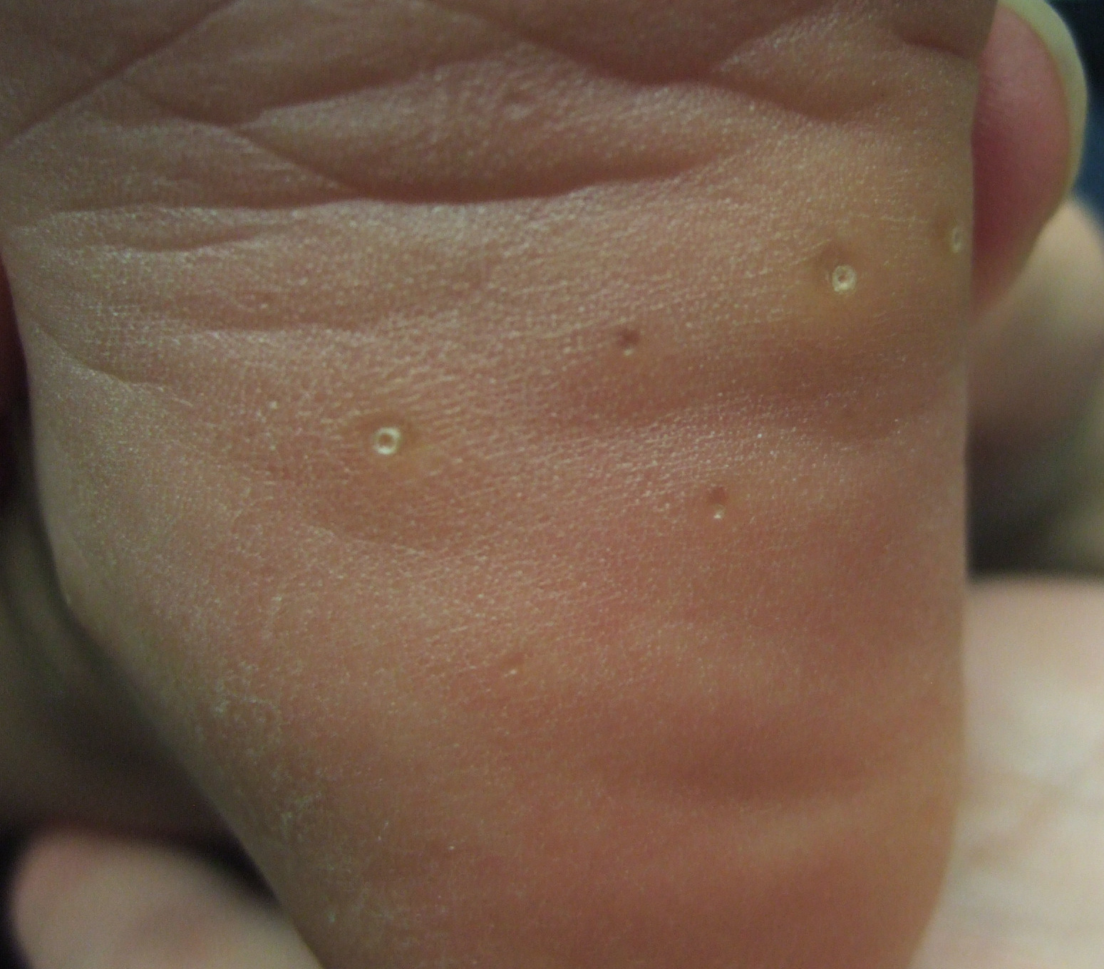
| Figure 1 | Figure 2 |
|---|
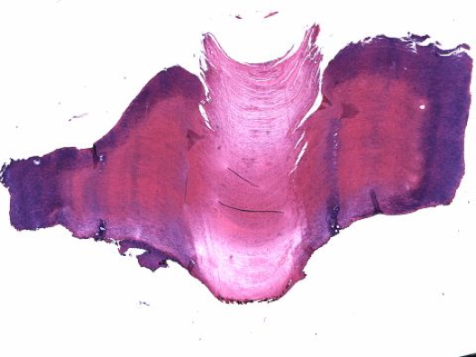 |
| Figure 3 |
|---|
A 2-year-old healthy Hispanic boy presents with a 1-year history of thick scaly circular “bumps” on the palms and soles. They are largely asymptomatic, but with occasional pruritus. The boy has met all of his developmental milestones and has no other physical complaints. There are no other family members affected with the same condition. Skin exam reveals scattered well demarcated, firm, hyperkeratotic papules with a central keratin plug that are distributed along both palms and soles. Some papules have a central depression (Figures 1 and 2). A punch biopsy of a papule is shown in Figure 3.
The clinical findings of discrete hyperkeratotic papules on the palms and soles with histological findings of a cornoid lamella support a diagnosis of punctuate porokeratotic keratoderma. This diagnosis has been reported in patients of all races. However, to our knowledge this is the youngest patient with punctate porokeratotic keratoderma to be reported in the literature. Previous reported ages of onset range from 12 to 74 years [1, 2].
Clinically, punctate porokeratotic keratoderma is characterized by multiple asymptomatic keratotic papules on the palms and soles and less frequently the dorsal and lateral surfaces of the fingers [3]. We believe punctate porokeratotic keratoderma to be part of a clinical spectrum characterized by hyperkeratotic papules on the palms and soles with an underlying cornoid lamella. In the literature, spiny keratoderma has often been used interchangeably with punctate porokeratotic keratoderma but clinically it appears different because of the projection of multiple spiny papules from the palms and soles, but they may be variants of the same condition [4].
Both sporadic and autosomal cases of punctate porokeratotic keratoderma have been reported [5, 6, 7]. Sporadic cases have been linked with malignancies such as bronchial or ovarian carcinoma [8]. Spiny keratoderma has been associated with systemic conditions including type IV hyperlipoproteinemia, autosomal dominant polycystic kidney disease, and chronic lymphocytic leukemia [9, 10]. Gene defects associated with autosomal dominant inheritance have not been characterized thus far.
Etiology for the disordered keratinization underlying punctate porokeratotic keratoderma is unclear but may involve hyperproliferation of basal keratinocytes below the keratotic plug [9]. Histology shows a broad column of parakeratosis with focally decreased granular layer consistent with a cornoid lamella. This characteristic finding allows for differentiation of this condition from most of other forms of palmoplantar keratoderma.
 |
| Figure 4 |
|---|
Treatment of punctate porokeratotic keratoderma is difficult. Available options include topical 5-FU, vitamin D derivatives, or salicylic acid followed by curettage. Our patient responded to urea 40 percent cream with resolution of the central keratin plug but not the underlying nodules (Figure 4).
© 2010 Dermatology Online Journal