Giant cell tumor of soft tissue on the thigh of a 40-year-old woman
Published Web Location
https://doi.org/10.5070/D33kt093nbMain Content
Giant cell tumor of soft tissue on the thigh of a 40-year-old woman
Victoria Nguyen BS, Carlos Garcia MD, Henry Haskell MD
Dermatology Online Journal 16 (3): 2
1. Department of Dermatology at the Oklahoma University Health Sciences Center, Oklahoma City, Oklahoma. carlos-garcia@ouhsc.edu2. Department of Pathology at the Oklahoma University Health Sciences Center, Oklahoma City, Oklahoma
Abstract
We report a case of giant cell tumor of soft tissue (GCTST) in a 40-year-old woman who presented with a painful fibrous nodule on the thigh. The histological examination revealed multinucleated histiocytes admixed with eosinophils, lymphocytes, and scattered spindle-shaped cells. The clinical presentations, histological features, differential diagnosis, treatment options, and a literature review are presented.
Case report
 |  |
| Figure 1 | Figure 2 |
|---|---|
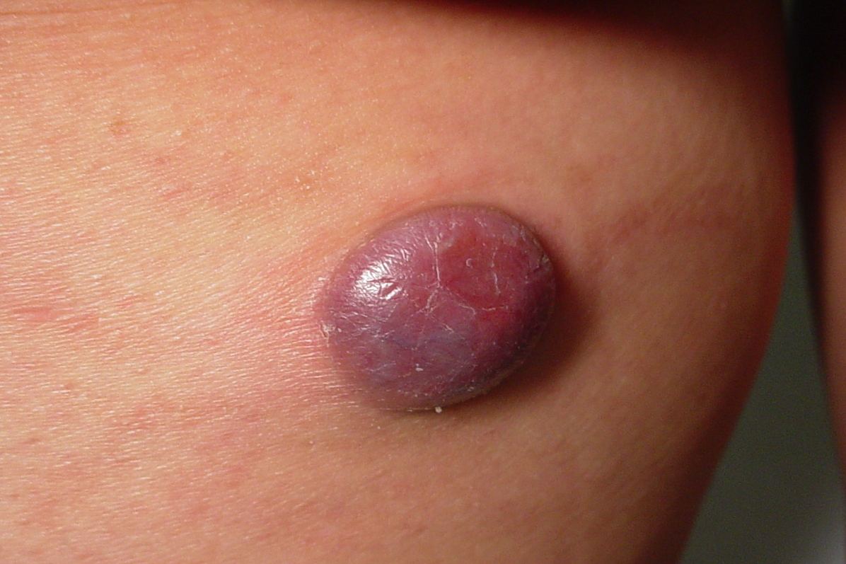
| Figure 1. A large, smooth, firm, and erythematous nodule on the posterior aspect of the left thigh. The lesion was not attached
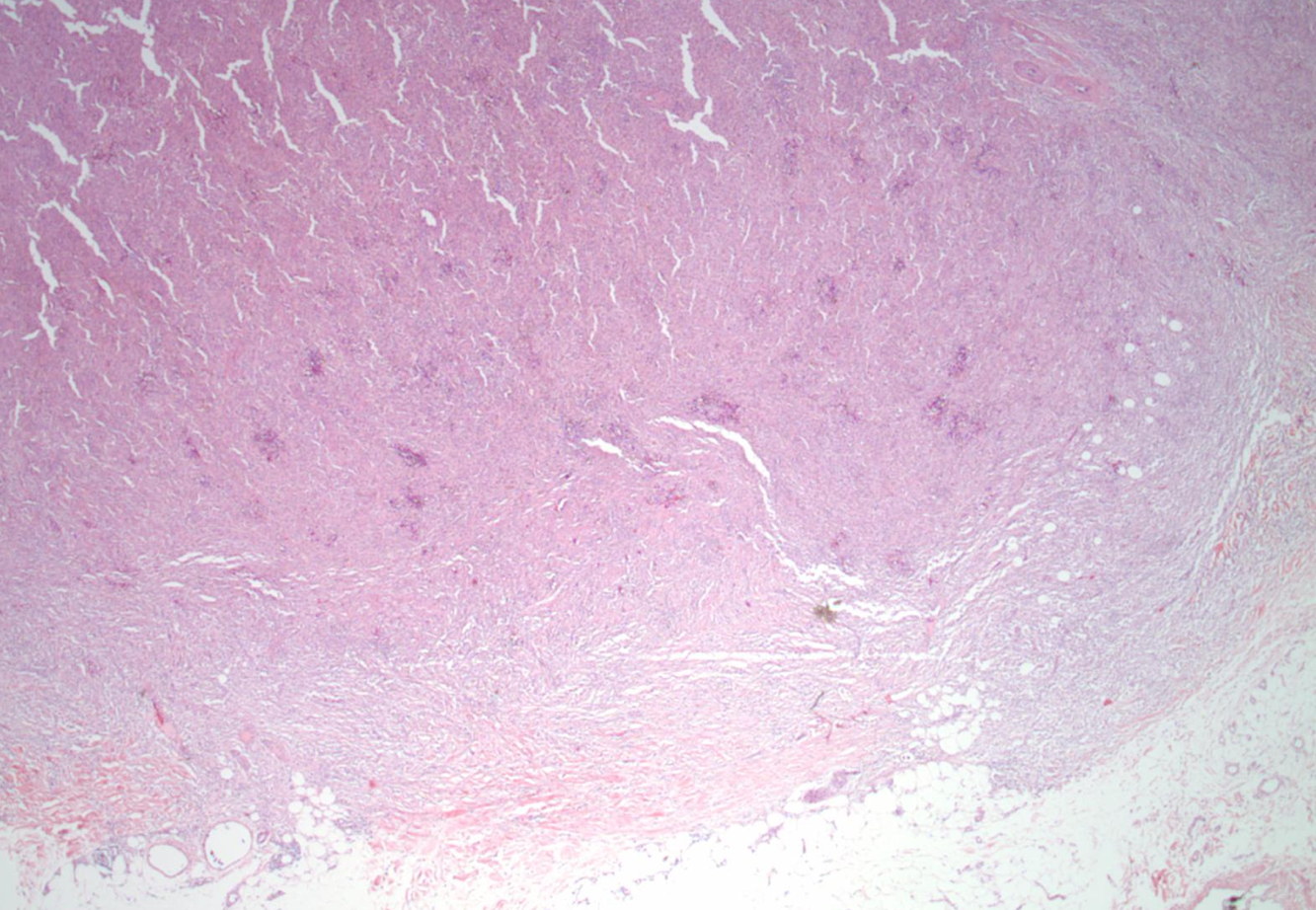
to deeper structures and was mildly tender on palpation. Figure 2. The sections show a large, well-defined tumor abutting the subcutis (x2). | |
AA 40-year-old woman with history of depression and stroke presented with a 3-month history of a growing painful nodule on her left posterior thigh. Physical examination revealed a 2 cm firm, smooth, well-demarcated, erythematous, violaceous, and mildly tender nodule (Figure 1). The lesion began spontaneously as a small red papule that grew rapidly.
The tumor was completely excised and sent for microscopic examination (Figures 2 through 4).
 |  |
| Figure 3 | Figure 4 |
|---|---|
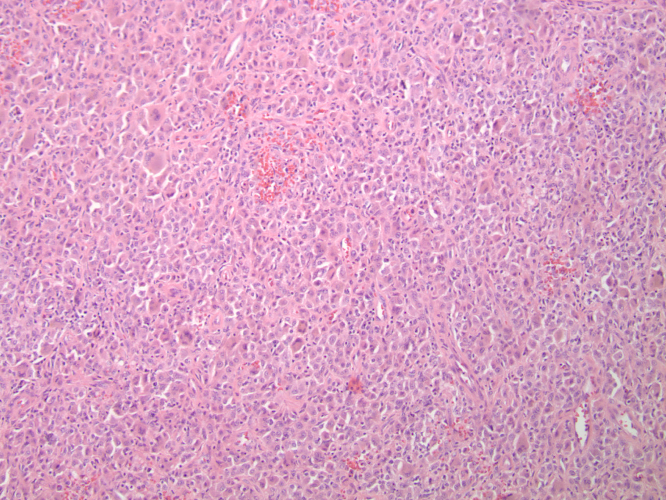
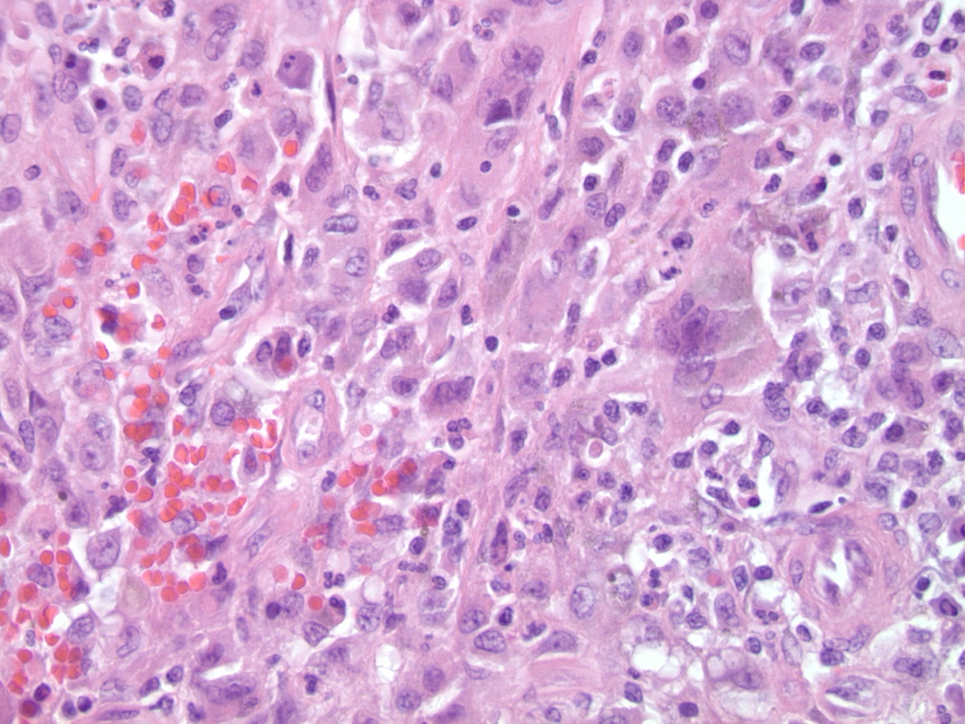
| Figure 3. There are numerous osteoclast-like giant cells, multiple foci of hemorrhage, and hemosiderin accumulation (x10). Figure 4. Stromal hemorrhage, hemosiderin deposition, and an osteoclast-like giant cell (x400). | |
Histological examination revealed a dermal tumor characterized by a round, well-circumscribed nodule of multinucleated histiocytes admixed with eosinophils, lymphocytes, and scattered spindle-shaped cells. Occasional mitoses but no fully transformed malignant change or atypical features were appreciated. There was no prominent bone formation at the periphery of the tumor. The morphological features are those of a giant cell tumor of soft tissue. The lesion was completely excised and the patient has remained recurrence-free for 23 months.
Discussion
Giant cell tumor of soft tissue, first described by Salm and Sissons in 1972, is a rare neoplasm located in both superficial and deep soft tissue [1]. Giant cell tumor of soft tissue most commonly involves the upper and lower extremities, but the trunk, head, and neck are not unusual sites. It often presents as a painless, firm, mobile, well-demarcated mass with no connection to the underlying muscle, tendon, or bone. There are no reported gross features that distinguish between benign and malignant variants. The few reported cases in the dermatologic literature are summarized in Table 1.
The hallmark of GCTST is a nodular arrangement of tumor cells distributed in a cellular fibroblastic stroma. The tumor is composed of a mixture of mononuclear and osteoclast-like giant multinucleated cells that closely resemble those found in the GCT of bone. The mononuclear cells have an epithelioid or spindled appearance, eosinophilic cytoplasm, and centrally located nuclei [2, 3]. A fascicular pattern with focal storiform arrangement of spindle mononuclear cells may be noted [4]. The malignant variant contains giant mononuclear cells with hyperlobated nuclei and mononuclear cells with conspicuous nuclear pleomorphism and hyperchromasia [3].
Immunohistochemically, GCTST stains diffusely positive for CD68 within multinucleated osteoclast-like cells. It stains focally positive for CD6, Ham 56, and smooth muscle actin within mononuclear cells [2, 5]. Vimentin and alpha-1-antichymotrypsin are positive in both osteoclast-like cells and mononuclear cells [4, 6]. Ki-67 has been found positive in mononuclear cells [6].
The differential diagnosis should include dermatofibroma, atypical fibroxanthomas, plexiform fibrohistiocytic tumor, giant cell malignant fibrous histocytoma, osteoclast-like giant cell rich leiomyosarcoma, extraskeletal osteosarcoma [4], nodular fascitis with giant cells, epithelioid sarcoma, and tenosynovial giant cell tumor. Most cases of GCTST follow a benign clinical course but can be locally aggressive and recur locally. Whereas metastases are infrequent, there have been reported cases of metastases to the lungs [6, 7] and parotid glands [8]. Superficial GCTST appears to have a more favorable prognosis than deep-seated GCTST. The treatment of choice for GCTST is complete surgical excision [1]. The reported recurrence rate is 6.2 percent [9].
References
1. Salm R, Sissons HA. Giant-cell tumors of soft tissues. J Pathol. 1972;107:27-39. [PubMed]2. May SA, Deavers MT, Resetkova E, Johnson D, Albarracin CT. Giant cell tumor of soft tissue arising in breast. Ann Diagn Pathol. 2007;11(5):345-9. [PubMed]
3. O'Connell JX, Wehrli BM, Nielsen GP, Rosenberg AE. Giant cell tumors of soft tissue: a clinicopathologic study of 18 benign and malignant tumors. Am J Surg Pathol. 2000;24(3):386-95. [PubMed]
4. Hoang MP, Rogers BB, Albores-Saavedra J. Giant cell tumor of the skin: a morphologic and immunohistochemical study of five cases. Ann Diagn Pathol. 2002;6(5):288-93. [PubMed]
5. Angervall L, Hagmar B, Kindblom LG, Merck C. Malignant giant cell tumor of soft tissues: a clinicopathologic, cytologic, ultrastructural, angiographic, and microangiographic study. Cancer. 1981;47(4):736-47. [PubMed]
6. Guo H, Garcia RA, Perle MA, Amodio J, Greco MA. Giant cell tumor of soft tissue with pulmonary metastases: pathologic and cytogenetic study. Pediatric Dev Pathol. 2005;8:718-24. [PubMed]
7. Oliveira AM, Dei Tos AP, Fletcher CD, Nascimento AG. Primary giant cell tumor of soft tissues: A study of 22 cases. Am J Surg Pathol. 2000;24:248-256. [PubMed]
8. Grabellus F, von Winterfeld F, Sheu SY, Metz KA, Jahnke K, Schmid KW. Unusual aggressive course of a giant cell tumor of soft tissue during immunosuppressive therapy. Virchows Arch. 2006;448(6):847-51. [PubMed]
9. Unni, KK. Dahlin's bone tumors: general aspect and data on 11087 cases. 5. Philadelphia: Lippincott-Raven; 1998.
© 2010 Dermatology Online Journal