Prurigo pigmentosa treated with doxycycline
Published Web Location
https://doi.org/10.5070/D39s7059nhMain Content
Prurigo pigmentosa treated with doxycycline
Tugba Rezan Ekmekci, Ilknur Kivanc Altunay, and Adem Koslu
Dermatology Online Journal 12 (1): 9
Departmant of Dermatology, Sisli Etfal Research and Training Hospital, Istanbul, TURKEY. tre@ttnet.net.trAbstract
Prurigo pigmentosa is characterized by an inflammatory phase with pruritic erythematous papules and a resolution phase with reticulated pigmentation. It is not a well known entity except in Japan. We present a Turkish young man with prurigo pigmentosa treated with doxycycline.
Prurigo pigmentosa (PP), first described by Nagashima in 1971, is an uncommon, inflammatory dermatitis of unknown etiology [1]. It is now regarded as a distinct clinical entity. Outside of Japan, it is very rare disorder [2].
We describe a Turkish young man with a typical clinical presentation and supportive histological findings of prurigo pigmentosa who was treated successfully with doxycycline.
Clinical synopsis
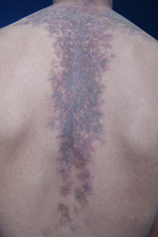
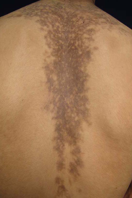
A 17-year-old Turkish man with skin type IV presented with a 5-month history of an intensely pruritic eruption on the upper back and chest. Despite therapy with topical corticosteroids and oral antihistamines, the eruption and the pruritus increased. His medical and family history were not contributory. Dermatogical examination revealed reticular brownish pigmentation with multiple erythematous, edematous, urticarial papules on the central region of his upper back (Fig. 1) and chest. These lesions exhibited scale, but there was no atrophy, telangiectasia, hypopigmentation, vesicles, or bullae. A biopsy specimen from erythematous papules showed spongiosis, exocytosis, edema, superficial perivascular neutrophilic and mononuclear infiltration. The biopsy from a brownish lesion revealed spongiosis, slight basal pigmentation, and perivascular-mononuclear infiltration, including a small number of melanophages in the dermal papillae and superficial reticular dermis. Results of laboratory investigations, including peripheral blood cell counts, blood glucose level, liver and renal function tests, and urinalysis were within normal limits. Standard patch tests gave negative results. He was diagnosed with prurigo pigmentosa with a typical clinical picture and a supportive biopsy. He was started on 100 mg of oral doxycycline daily for 4 weeks without topical treatment. After 10 days, the pruritus and edematous papules disappeared leaving reticular brownish pigmentation (Fig. 2). No recurrence was observed within the following 6 months.
 |  |
| Figure 1 | Figure 2 |
|---|---|
| Figure 1: Reticular brownish pigmentation with erythematous, edematous, urticarial papules and scale distributed symmetrically and in wedge shape on the upper back before treatment. | |
| Figure 2: The eruption was markedly improved after a 10-day treatment with doxycycline, leaving marked reticular pigmentation. | |
Discussion
Prurigo pigmentosa is a recurrent pruritic eruption characterized by the sudden onset of erythematous papules that coalesce to form a reticulate pattern. The papules may evolve into urticarial plaques and may develop scale. The resolution of the primary lesions occurs within days, leaving a mottled or reticulate hyperpigmentation [3]. The distribution of the lesions is on the back, including the upper back and scapular regions, nucha, clavicular regions, and chest. However, the lesions may occur on other sites, such as the abdomen, lumbosacral regions, antecubital fossae, limbs, and forehead. The mucous membranes are spared.
Total duration may range from 6 months to 8 years. Despite a chronic course, lichenification of the lesions is surprisingly rare [3]. The female:male ratio is 2:1 [4]. Prurigo pigmentosa is seen mostly in the spring and summer [5]. The lesion of our case had commenced during the spring.
The pathogenesis of PP remains unclear [2]. The frequency of the initial cases from Japan suggested an ethnic predisposition or environmental cause [6]. Several hypothesis about the mechanism of this disease have been proposed, including friction with clothes or allergic reaction [2, 3]. Recent reports have demonstrated the association of this disease with systemic conditions such as diabetes mellitus, ketosis, pregnancy, and fasting or dieting [2, 7]. Any associated disease or a clue to its etiopathogenesis was absent in the presented case.
Prurigo pigmentosa begins with a superficial perivascular infiltrate of neutrophils. Shortly thereafter, neutrophils are scattered in dermal papillae and then sweep rapidly through an epidermis accompanied by spongiosis, ballooning, and necrotic keratinocytes. Soon thereafter, eosinophils and lymphocytes come to predominate over the neutrophils in a dermal infiltrate that assumes a patchy lichenoid pattern [4]. Epidermal changes consist of parakeratosis, exocytosis of lymphocytes, spongiosis, and liquefaction degeneration [8].
The differential diagnosis of PP includes lichen pigmentosus, pigmented contact dermatitis, confluent and reticulate papillomatosis, and erythema dischromicum perstans [5, 9]. However, these conditions are easily differentiated from PP by histology and clinical findings, such as recurrent papules and pruritus with sudden onset [9].
Topical and systemic corticosteroids or antihistamines are not beneficial in PP [4]. Nowadays, therapy with oral dapsone, sulfamethoxazole or minocycline is considered as standard [2]. But these drugs do not lessen the pigmentation in reticular pattern.
Dapsone and minocycline inhibit migration and function of neutrophils. At an incipient stage of the disease when neutrophils are preponderant, administration of these drugs may abort the pathologic process [8]. Macrolide antibiotics have recently been demonstrated to be beneficial in patients with PP who were previously unresponsive to minocycline [2]. Doxycycline has been reported to be effective in the treatment of PP [5, 6, 10]. We also treated our patient successfully with doxycycline.
References
1. Nagashima M. Prurigo pigmentosa:clinical observations of our 14 cases. J Dermatol. 1978 Apr;5(2):61-7. PubMed2. Yazawa N, Ihn H, Yamane K, Etoh T, Tamaki K. The successful treatment of prurigo pigmentosa with macrolide antibiotics. Dermatology. 2001;202(1):67-9. PubMed
3. Kim MH, Choi YW, Choi HY, Myung KB. Prurigo pigmentosa from contact allergy to chrome in detergent. Contact Dermatitis. 2001 May;44(5):289-92. PubMed
4. Boer A, Misago N, Wolter M, Kiryu H, Wang XD, Ackerman AB. Prurigo pigmentosa: a distinctive inflammatory disease of the skin. Am J Dermatopathol. 2003 Apr;25(2):117-29. PubMed
5. Gur-Toy G, Gungor E, Artuz F, Aksoy F, Alli N. Prurigo pigmentosa. Int J Dermatol. 2002 May;41(5):288-91. PubMed
6. Erbagci Z. Prurigo pigmentosa in association with Helicobacter pylori infection in a Caucasian Turkish woman. Acta Derm Venereol. 2002;82(4):302-3. PubMed
7. Yokozeki M, Watanabe J, Hotsubo T, Matsumura T. Prurigo pigmentosa disappeared following improvement of diabetic ketosis by insulin. J Dermatol. 2003 Mar;30(3):257-8. PubMed
8. Boer A, Ackerman AB. Prurigo pigmentosa is distinctive histopathologically. Int J Dermatol. 2003 May;42(5):417-8. PubMed
9. Matsumoto C, Kinoshita M, Baba S, Suzuki H, Kanematsu S, Kanematsu N. Vesicular prurigo pigmentosa cured by minocycline. J Eur Acad Dermatol Venereol. 2001 Jul;15(4):354-6. PubMed
10. Gurses L, Gurbuz O, Demircay Z, Kotiloglu E. Prurigo pigmentosa. Int J Dermatol. 1999 Dec;38(12):924-5. PubMed
© 2006 Dermatology Online Journal