Subacute cutaneous lupus erythematosus (SCLE) presenting in childhood
Published Web Location
https://doi.org/10.5070/D34g28d2dxMain Content
Subacute cutaneous lupus erythematosus (SCLE) presenting in childhood
Vandana Mehta Rai MD, and C Balachandran MD
Dermatology Online Journal: 11 (2): 27
Department of Skin and Sexually Transmitted Diseases, Kasturba Medical College,Manipal Karnataka. vandanamht@yahoo.com
Abstract
A 13-year-old boy presented with erythematous scaly plaques on the face and extremities with history of photosensitivity of 7-years duration. There was no history of oral ulcers or joint pains. Although the histopathological findings were inconclusive, a positive lupus band test confirmed the diagnosis of subacute cutaneous lupus erythematosus.
Introduction
Subacute lupus eythematosus (SCLE) is a distinct subset of cutaneous lupus characterised clinically by psoriasiform and or annular lesions. SCLE is extremely rare in childhood. We report a case of a 13-year-old boy who developed skin lesions suggestive of SCLE.
Clinical synopsis
|
|
| Figure 1 | Figure 2 |
|---|---|
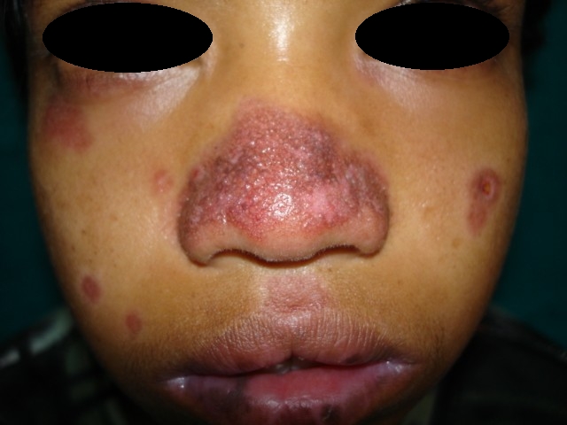
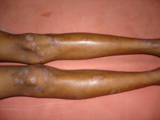
| Close view of the face. Lower Limbs | |
A 13-year-old boy presented with complaints of erythematous lesions on the body of 7-years duration. The child was born of a nonconsanguinous marriage and had normal growth and developmental milestones. At the age of 6 years he started developing erythematous papules on the nose; these coalesced to form scaly plaques. He developed similar lesions on the forehead, malar area, chin, and ears.
|
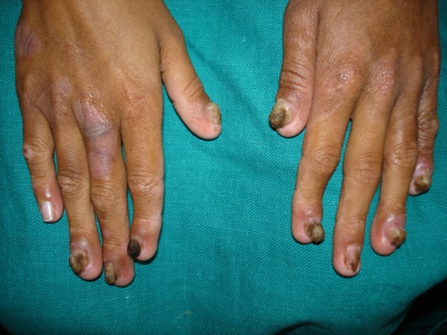
| Figure 3 |
|---|
| Nail dystrophy |
There was history of associated pruritus with photosensitivity, however, there was no history of oral ulcers or joint pains. At the same time he developed erythematous scaly plaques on the elbows and knees and thick scale on the scalp. Cutaneous examination revealed erythematous scaly plaques on the bridge of nose, malar area, chin, forehead, and ears. He had erythematous scaly hyperkeratotic plaques on the elbows and knees. His scalp showed thick adherant scale. Palms, soles, and the oral cavity were normal, however, the fingernails and toenails showed dystrophy. Based on the history and clinical findings a differential diagnosis of psoriasis or SCLE was considered and patient was further evaluated. Routine investigations such as hemogram, urinanalysis, LFT, RFT, C3 C4, ASO titre, and ANA profile were normal. The ESR was raised (65/hr). Direct immunofluorescence (DIF) from facial lesion showed a granular linear IgG and a discontinuous IgM band at the basement menbrane zone. DIF from the uninvolved skin of forearm was negative. A biopsy for histopathology from the leg lesion showed psorasiform hyperplasia with perivascular mononuclear infiltrate.
Discussion
SCLE was first described as a distinct subset of lupus erythematosus in 1979 by Sontheimer et al. [1]. Patients with SCLE present with erythematous, nonscarring papulosquamous or annular polycyclic lesions in a photosensitive distribution with scale, pigment changes, and telangiectasia associated with mild systemic symptoms (the most prominent of which are musculoskeletal complaints). Antinuclear antibodies are frequently present and patients produce anti-Ro antibodies. Although the classic presentation is in the form of annular or papulosquamous lesions, poikilodermatous [2], erythrodermic [3], and bullous SCLE [4] have also been reported. SCLE has a distinct histopathology with epidermal atrophy, hydropic degeneration of basal cell layer, and perivascular infiltrate in the dermis. Compared to DLE where over 90 percent of patients have immunoreactants at the dermoepidermal junction, 40-50 percent of SCLE patients do not show immune deposits. Approximately one half of SCLE patients fulfil the criteria for lupus erythematosus as per American Rheumatism Association (ARA) criteria [5]. Our patient was diagnosed as SCLE on the basis of nonscarring erythematous scaly plaques on face with history of photosensitivity. Although the histopathology results were inconclusive a positive lupus band test confirmed the diagnosis. SCLE is extremely rare in childhood though a few cases have been reported in literature [6, 7, 8]. There have been no reports of SCLE in childhood from India to our knowledge. Another unusual feature in our case was also the nail dystrophy affecting almost all the nails which has not been reported earlier. Our patient improved with potent topical corticosteroids and oral hydroxychloroquin.
References
1. Sontheimer RD, Thomas JR, Gillian JN. SCLE- a cutaneous marker for a distinctive LE subset. Arch Dermatol 1979;115:1409-4152. Marzano AV, Facchetti M, Alessi E. Poikilodermatous Subacute cutaneous lupus erythematosus. Dermatology 2003;207:285-90
3. Mutasim DF. Severe subacute cutaneous lupus erythematosus presenting with generalised erythroderma and bullae. J Am Acad Dermatol 2003;48:947-9
4. Perera GK, Black MM, Mc Gibbon DH. Bullous Subacute cutaneous lupus erythematosus. Clin Exp Dermatol 2004;29:265-7
5. Herrero C, Bielsa J, Font J. Subacute cutaneous lupus erythematosus. Clinical pathological findings in 13 cases. J Am Acad Dermatol 1988;19:1057-62
6. Ciconte A, Mills AE, Shipley A, Marks R. Subacute cutaneous lupus erythematosus presenting in a child. Australas J Dermatol 2002;43:62-4
7. Siamopoulou-Mavridou A, Stefanou D, Drosos SS. Subacute cutaneous lupus erythematosus in childhood. Clin Rheumatol 1989;8:533-7
8. Amato L, Coronella G, Berti S, Moretti S, Fabbri P. Subacute cutaneous lupus erythematosus in childhood Pediatr Dermatol 2003;20:31-4
© 2005 Dermatology Online Journal